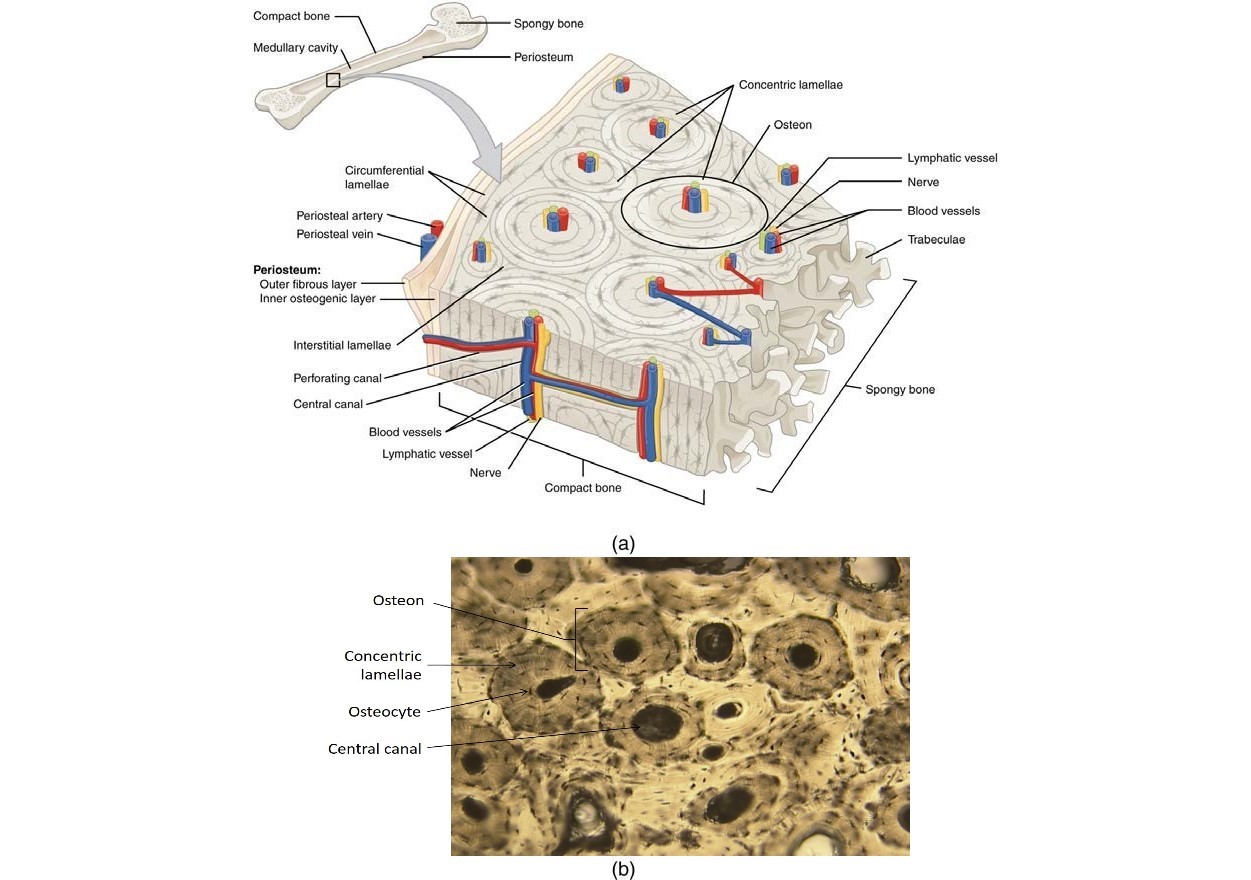
Compact Bone Diagram : Structure of compact bone. (a) Cross-sectional view of ... : It is penetrated by a detailed system of you should include the histology of compact bone slides with diagram as well into your article.
Compact Bone Diagram : Structure of compact bone. (a) Cross-sectional view of ... : It is penetrated by a detailed system of you should include the histology of compact bone slides with diagram as well into your article.. Mature compact bone is structurally layered or lamellar. A typical long bone showing gross anatomical features. Compact bone diagram osteon compact bone ap pinterest anatomy human anatomy and. The three types of cartilages 1. Schematic diagram for cross and longitudinal sections of long bone showing the compact bone formed from osteons that are consisted of circumferential bone lamellae around the haversian canals.
They are the bones of your forearm. Compact bone diagram osteon compact bone ap pinterest anatomy human anatomy and. Compact bone, also known as cortical bone, is a denser material used to create much of the hard structure of the skeleton. Cortical bone contains haversian systems (osteons) which contain a central haversian canal surrounded by osseous tissue in a. Cortical bone forms the extremely hard exterior while cancellous bone fills the interior.
TERRY ANATOOMY: movement from photos1.blogger.com Compact bone diagram osteon compact bone ap pinterest anatomy human anatomy and. What are the 2 main types of bone? Compact bone is part of a bone made of densely packed tissue. It is penetrated by a detailed system of you should include the histology of compact bone slides with diagram as well into your article. Cortical bone is compact bone, while cancellous bone is trabecular and spongy bone. Compact bone high resolution histology diagram. Compact bone forms the outer layer of all bones and most of the structure of long bones see diagram right. A typical long bone showing gross anatomical features.
The outer walls of the diaphysis cortex cortical bone are composed of dense and hard compact bone a form of osseous tissue.
Long bones, like the tibia and fibula, are those bones whose. The worksheets are offered in developmentally. The two types of bones are compact bones and spongy bones. A diagram of the anatomy of a bone, showing the compact bone. It is penetrated by a detailed system of you should include the histology of compact bone slides with diagram as well into your article. Feel free to use for study purposes. The three types of cartilages 1. Click on the image to enlarge it. Cortical bone forms the extremely hard exterior while cancellous bone fills the interior. Compact bone diagram bone cross section diagram file624 diagram of compact bone new. Compact bone high resolution histology diagram. Label compact and spongy bone illustrations as demonstrated in class. What are the 2 main types of bone?
Compact bone high resolution histology diagram. The worksheet is an assortment of 4 intriguing pursuits that will enhance your kid's knowledge and abilities. Other sets by this creator. The three types of cartilages 1. The worksheets are offered in developmentally.
5.4: Bone Structure - Medicine LibreTexts from med.libretexts.org Compact bone diagram osteon compact bone ap pinterest anatomy human anatomy and. As seen in the image below, compact bone forms the cortex, or hard outer. The inner surface of compact bone is lined by a thin, cellular layer. Feel free to use for study purposes. What are diplo , its function, and location? Compact bone diagram osteon compact bone ap pinterest anatomy human anatomy and. Label compact and spongy bone illustrations as demonstrated in class. Schematic diagram for cross and longitudinal sections of long bone showing the compact bone formed from osteons that are consisted of circumferential bone lamellae around the haversian canals.
Label bone diagram s are getting used for different functions from past many years.
Label compact and spongy bone illustrations as demonstrated in class. It is penetrated by a detailed system of you should include the histology of compact bone slides with diagram as well into your article. Label compact and spongy bone illustrations as demonstrated in class. What are diplo , its function, and location? A typical long bone showing gross anatomical features. Feel free to use for study purposes. Spongy bone is composed of trabeculae that contain the. You may also save it to your computer for more zoomed view. What are the 2 main types of bone? The radius is the bone which is present laterally, which mean. Cortical bone is compact bone, while cancellous bone is trabecular and spongy bone. The worksheets are offered in developmentally. Cortical bone forms the extremely hard exterior while cancellous bone fills the interior.
Feel free to use for study purposes. The outer shell of compact bone is called cortical bone or cortex. The three types of cartilages 1. What are diplo , its function, and location? Microscopic anatomy of compact bone.
Compact and Spongy Bone | Microanatomy of bone tissue ... from i.pinimg.com The worksheet is an assortment of 4 intriguing pursuits that will enhance your kid's knowledge and abilities. Label bone diagram s are getting used for different functions from past many years. The outer walls of the diaphysis cortex cortical bone are composed of dense and hard compact bone a form of osseous tissue. They are the bones of your forearm. I'm not sure of what you mean by bone diagram. Like compact bone, spongy bone, also known as cancellous bone, contains osteocytes housed in lacunae, but they are not arranged in concentric circles. Cortical bone forms the extremely hard exterior while cancellous bone fills the interior. Feel free to use for study purposes.
Feel free to use for study purposes.
The worksheet is an assortment of 4 intriguing pursuits that will enhance your kid's knowledge and abilities. Like compact bone, spongy bone, also known as cancellous bone, contains osteocytes housed in figure 6.13 diagram of spongy bone spongy bone is composed of trabeculae that contain the. Reader view spongy bone compact bone Feel free to use for study purposes. Long bones, like the tibia and fibula, are those bones whose. The radius and ulna are two parallel. Cortical bone is compact bone, while cancellous bone is trabecular and spongy bone. I'm not sure of what you mean by bone diagram. Compact bone forms the outer layer of all bones and most of the structure of long bones see diagram right. Label compact and spongy bone illustrations as demonstrated in class. Schematic diagram for cross and longitudinal sections of long bone showing the compact bone formed from osteons that are consisted of circumferential bone lamellae around the haversian canals. The radius is the bone which is present laterally, which mean. Compact bone, also known as cortical bone, is a denser material used to create much of the hard structure of the skeleton.


0 Komentar